<iframe src="https://www.googletagmanager.com/ns.html?id=GTM-W3R8R6G&gtm_auth=AU1ugrxhJK_EQcp9wg5qxA&gtm_preview=env-1&gtm_cookies_win=x" height="0" width="0" style="display:none;visibility:hidden"></iframe>

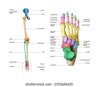
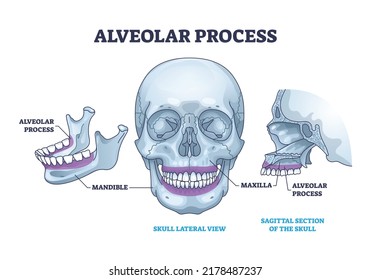
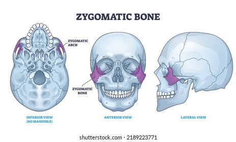
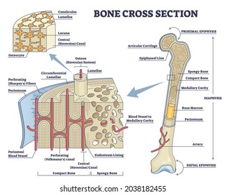
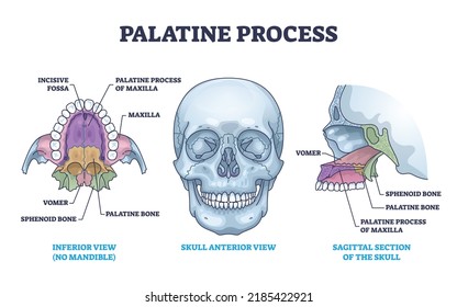
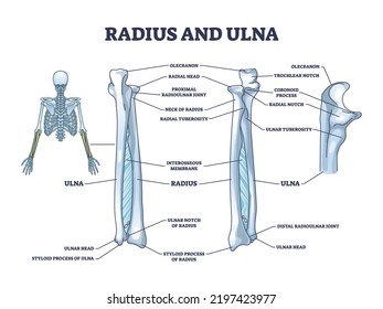
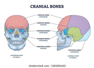
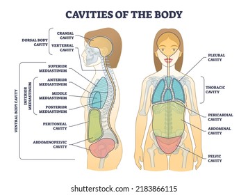
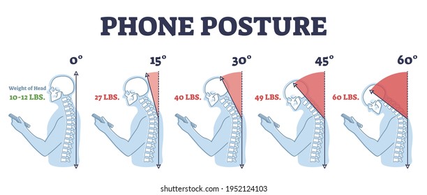
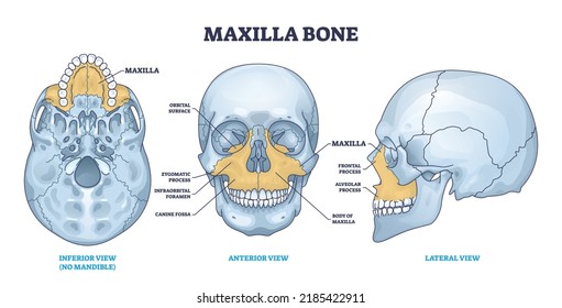
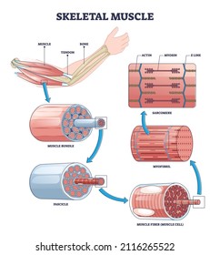
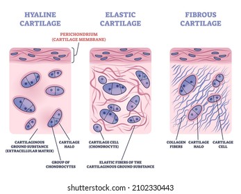
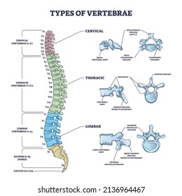
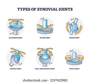
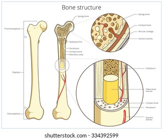
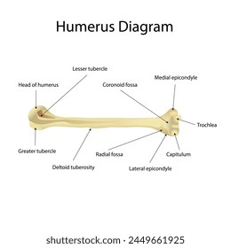
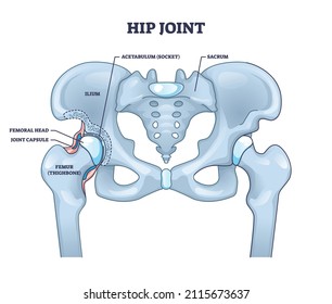
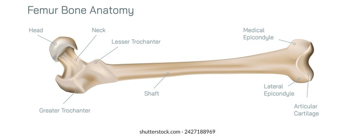
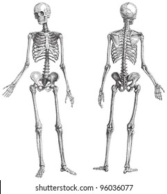
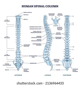
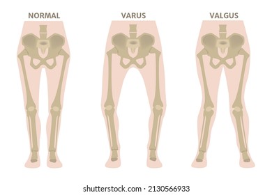
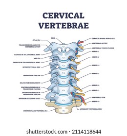
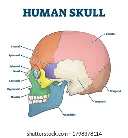
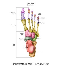
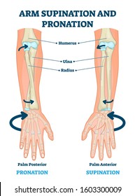
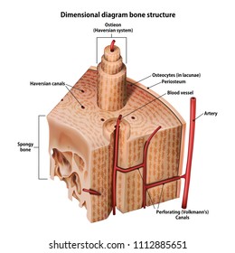
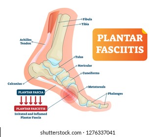
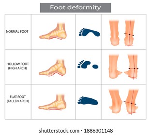
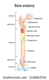
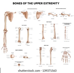
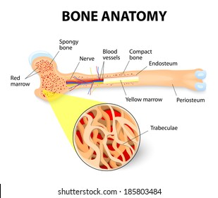
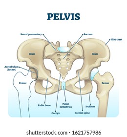
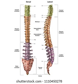
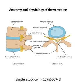
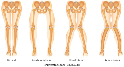
45 bone anatomy designs graphics for t-shirt and print on demand merch

Download bone anatomy t-shirt designs and other merch graphics like book covers, phone cases, tote bags and more.

Sort by

Most relevant
Diagram of the structure of the bones of a human leg on a white background. Vector 3D illustration
Alveolar process with anatomical head bone ridge for teeth outline diagram. Labeled educational scheme with chin maxilla and mandible parts vector illustration. Dental implant location on human skull.
Zygomatic bone location with human skull skeleton anatomy outline diagram. Labeled educational scheme with cheekbone from inferior, anterior and lateral view vector illustration. Head parts
Bone cross section and isolated anatomical detailed structure outline diagram. Labeled educational medical body description with distal and proximal epiphysis and osteon closeup vector illustration.
Palatine process section anatomy with maxilla structure outline diagram. Labeled educational facial skeletal bone description with incisive fossa, vomer, sphenoid and palatine vector illustration.
Radius and ulna bone anatomy with arm skeletal structure outline diagram. Labeled educational scheme with upper body parts and hand long bones vector illustration. Detailed physiological description.
Cranial bones anatomy and skull skeleton medical division outline diagram. Labeled educational frontal, parietal, sphenoid, ethmoid and temporal bone vector illustration. Paired and unpaired examples.
Cavities of body and anatomical compartment medical division outline diagram. Labeled educational scheme with physical dorsal, ventral and inferior mediastinum location explanation vector
Structure of muscle with isolated myosin and actin closeup outline diagram. Labeled educational arm bone muscular inner parts detailed description with sarcomere magnification vector illustration.
Anterior pelvic tilt or APT as pelvis abnormal posture outline diagram. Labeled educational scheme with syndrome from increased lordosis of lumbar spine and protrusion of abdomen vector illustration.
The anatomy of the human foot is medial and middle plantar muscles. Vector 3D illustration
Growing bones vector illustration. Educational fetus, childhood and adolescence organ development stages scheme. Healthy lifetime process infographic. Ossification center and spongy formation graph
density of a healthy bone. Cross section of human femur. Inside of a bone showing the trabecular structure. Close-up of bone tissue. looking through a magnifying glass. Vector illustration
Spongy vs compact bone comparison with anatomical structure outline diagram. Labeled educational skeletal structural differences with porous inner and rigid outer diaphysis parts vector illustration.
Vector illustration of human long bone anatomy. Cross-section of a long bone showing labeled internal structures.
Phone posture while standing for correct spine and neck angle outline diagram. Healthy smartphone browsing from anatomical view vector illustration. Head weight problem and potential medical injury.
Maxilla bone detailed structure and facial skeleton anatomy outline diagram. Labeled educational scheme with medical inferior view without mandible, anterior and lateral skull view vector
Skeletal muscle structure layers with anatomical arm closeups outline diagram. Labeled educational descriptions with bundle, fascicle, fibers, myofibril and sarcomere sections vector illustration.
Skeletal muscle description with cross section structure outline diagram. Labeled educational epimysium, perimysium and endomysium location with anatomical muscular explanation vector illustration.
Perichondrium as hyaline, fibrous and elastic cartilage membrane outline diagram. Labeled educational tissue layer structure with cartilaginous ground substance, halo and collagen vector illustration.
Types of vertebrae and cervical, thoracic and lumbar division outline diagram. Labeled educational scheme with spinal skeletal bones vector illustration. Human anatomy and backbone medical description
Femer Structure Science Design Vector Illustration Diagram
Types of synovial joints movement classification for body outline diagram. Labeled educational anatomical division with ellipsoid, hinge, saddle, pivot and ball socket bone joints vector illustration.
Bone structure medical educational science vector illustration. Bone anatomy
Meninges as central brain part structure or under skin layers outline diagram. Labeled educational and anatomical parts location scheme with healthy medical skull elements example vector illustration.
Humerus Labeled Diagram, rib diagram, Human Rib diagram
Hip joint structure with anatomical bone parts description outline concept. Labeled educational scheme with human sacrum, ilium, femoral head, joint capsule and femur thighbone vector illustration.
Types of bone fracture. Structure of a bone vector illustration. The quality or structure changes in marrow density. Build and strength of bone tissues. Stymptoms and stages and  growth. Bone anatomy.
Human skeleton (male) / vintage illustration from Meyers Konversations-Lexikon 1897
Bones of the human hand with the name and description of all sites. Human anatomy. Vector illustration isolated on a white background.
Human spinal column structure with backbone and spine skeletal anatomy outline diagram. Labeled educational human back description and detailed anterior, lateral or posterior view vector illustration.
Valgus and varus leg deformities. Diagram showing the deformed bones of the lower extremities. Cosmetic pathology. Vector illustration.
Cervical vertebrae with bones detailed and labeled structure outline diagram. Educational anatomical back scheme with skeletal, nerves and artery in backbone and spinal section vector illustration.
Human skull bones skeleton labeled educational scheme vector illustration. Anatomical head zones separated with colors with side view diagram. Medicine study handout with parts title description.
Bones of the human foot with the name and description of all sites. Superior view. Human anatomy. Vector illustration isolated on a white background.
Arm supination and pronation vector illustration. Labeled anatomical scheme. Medical diagram with inner bones and joints. Compared palm posterior and anterior. Hand rotation movement biological terms.
Three-dimensional diagram bone structure
Vector illustration of flat feet vs normal feet with anatomical comparison, useful for podiatry education, medical presentations, orthopedic posters, and health awareness materials.
Plantar fasciitis vector illustration. Labeled human feet sport disorder diagram. Educational medical scheme with orthopedic leg disease. Painful plantar fascia inflammation and irritation infographic
Foot deformation. Types pathologies of foot. Hollow, flat and normal foot. Medical infographic.
Bone anatomy. Structure of a Long Bone. vector illustration for medical, educational and science use
Hallux valgus. Anatomy. Human foot bones. Signatures, also for clinics
Clavicle (collar bone), scapula (shoulder blade), humerus, ulna, radius, finger and hand. Detailed medical illustrations. Latin medical terms. Isolated on a white background.
bone remodeling process (resorption, reversal, formation, and mineralization). Osteoblast, osteoclast, and osteocyte. Vector illustration of human bone cell types. Medical diagram.
anatomy of the Long Bone. Periosteum, endosteum, bone marrow and trabeculae.
Pelvis anatomical skeleton structure. labeled vector illustration diagram. Medical education scheme with ilium, ischium, coccyx, sacrum, femur and pubic bone examples. Healthy human hips model image.
Illustration of the human spine with the name and description of all sites. Lateral and spinal views.
Anatomy and physiology of the vertebrae. Human vertebrae in superior and lateral views with main parts labeled. Vector illustration
Knee joint isolated on blue. Bone health, physiotherapy care, joint disorder, glucosamine repair, orthopedic surgery, physical therapy, magnesium supplement, osteoarthritis treatment concept
Bandy-legs. Shape of the legs - Genu varum, genu valgum - a physical legs deformity, bowing of the leg in relation to the thigh. Knock Knees - Valgus, is a physical deformity
Sponsored results by Shutterstock Logo
Get 15% off with code: VEXELS15